-
00:00
1.
Paraffin embedded tissue
-
00:03
1.1
Deparaffinized
-
00:23
1.2
Dehydrate
-
00:41
1.3
Air dry
-
00:52
1.4
Pre-treatment
-
01:24
1.5
Protease treatment
-
02:04
1.6
Dehydrate (Room temperature)
-
02:32
1.7
Air dry
-
02:34
2.
FISH protocol
-
02:36
2.1
Mark hybridizing area
-
02:42
2.2
Apply 10μl FISH probe for 22mm x 22mm area
-
02:54
2.3
Cover with cover glass and seal with rubber cement
-
03:27
2.4
Denature
-
03:36
3.
Incubation
-
03:55
4.
Remove rubber cement Slide into 2X SSC and remove cover glass
-
04:33
5.
Counter stain
-
04:35
5.1
Apply 10μl DAPI Solution to target area
-
04:43
5.2
Put on cover glass Seal with manicure
-
04:51
6.
Examine
播放影片: http://llai.cm.ntu.edu.tw/media/77
( http://www.abnova.com ) - FISH is a technique used to identify and localize the presence or absence of specific DNA sequences on cells and tissues. You can use FISH probes for the detection of gene amplification, loss and translocation. Each FISH probe product has a pair of locus-specific, fluorophore-labeled probes originated from a bacterial artificial chromosome (BAC) library.

附件:
重點
- 材料準備
- 1.Preparation of FISH probe1. The following FISH probes are ready-to-use, no need of any preparation.a. Gene FISH Probe (Cat # FGxxxx)
b. Split FISH Probe (Cat # FSxxxx)
c. Translocation FISH Probe (Cat # FTxxxx)
d. Prenatal FISH Probe (Cat # FMxxxx)
e. Made to Order FISH Probe (Ca # FAxxxx)2. Chromosome FISH Probe (Cat # FCxxxx) and Subtelomere FISH Probe
(Cat # FExxxx) are provided in 5x concentrated format, they should be either:a. Diluted to 1x with FISH Hybridization Buffer (Cat # U0028 or U0029) before use,
OR
b. Mixed with same category of FISH Probes (up to 5 different probes) to use, for example:- Combine 2 different probes:
1 volume of probe 1 (2 uL) + 1 volume of probe 2 (2 uL)
+ 3 volume of FISH Hybridization Buffer (6 uL) - Combine 3 different probes:
1 volume of probe 1 (2 uL) + 1 volume of probe 2 (2 uL)
+ 1 volume of probe 3 (2 uL) + 2 volume of FISH Hybridization Buffer (4 uL)
- Combine 2 different probes:
- 2.Recommended filter setThe table below is a recommendation of filter set use:
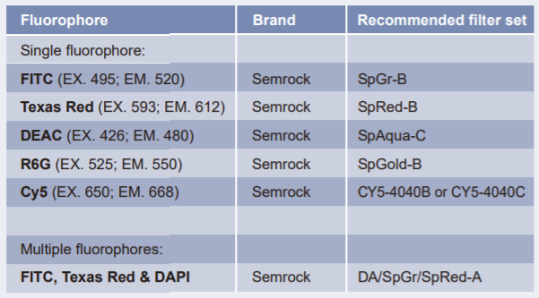 Note: EX. = excitation wavelength; EM. = emission wavelength
 - 3.Protocol selectionPlease follow an appropriate protocol below depend on the sample use, these samples include Paraffin embedded tissue (or FFPE), Frozen tissue and Metaphase spreads.For Paraffin embedded tissue, we recommended FFPE FISH PreTreatment Kit 1(Catalog #: KA2375 or KA2691 for the pretreatment of Formalin-Fixed Paraffin-Embedded (FFPE) tissue sections.
- 實驗步驟Paraffin embedded tissue
- 4.DeparaffinizedXylene 5 min×3 Room temperature
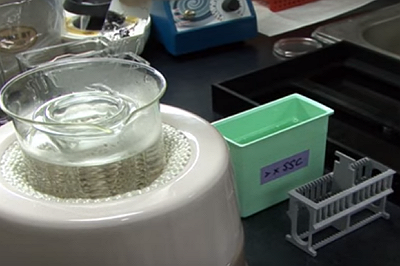
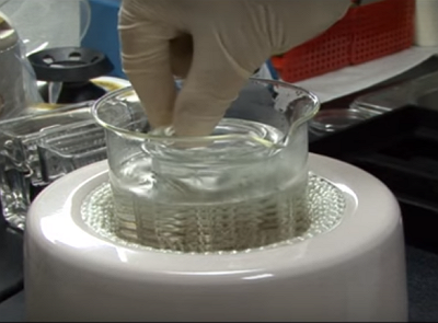
- 5.Dehydrate100% EtOH 5 min×2 Room temperature
- 6.Air dry

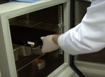
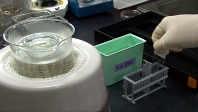
- 7.Pre-treatmentParaffin Pretreatment Solution 95℃ 30 minWash buffer (2×SSC) 5 min×2

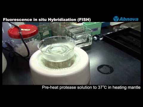
- 8.Protease treatmentProtease Solution 37℃ 10∼20min
Wash buffer (2×SSC) 5 min×2
NOTE:
*Protease Solution Add 500μl protease in 50ml protease buffer
*Protease preservation One month:4℃ Over one month:-20℃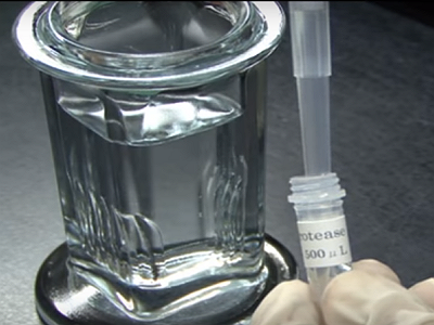
- 9.Dehydrate (Room temperature)70% EtOH 1 min100% EtOH 1 min
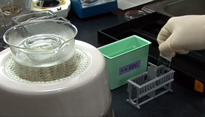
- 10.Air dry
- FISH protocol
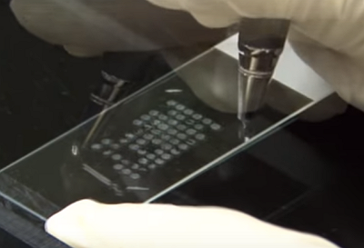
- 11.Mark hybridizing areause diamond pen
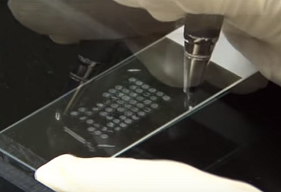
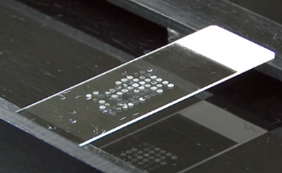
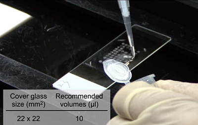
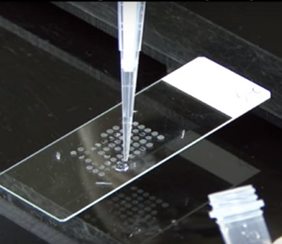
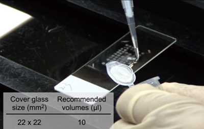
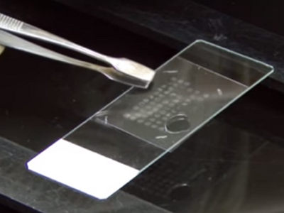
- 12.Apply 10μl FISH probe for 22mm x 22mm area

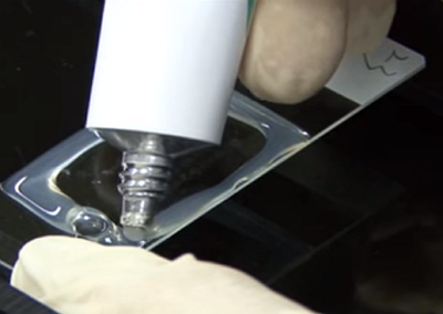
- 13.Cover with cover glass and seal with rubber cement

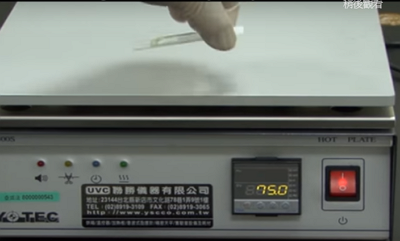
- 14.Denature75℃ 5 min

- Hybridization
- 15.IncubationHumidified box 37℃ 16 ~ 72 hrs
- Wash procedure
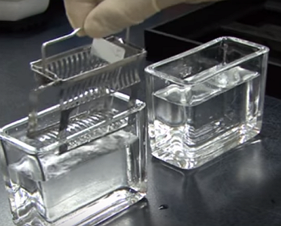
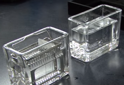
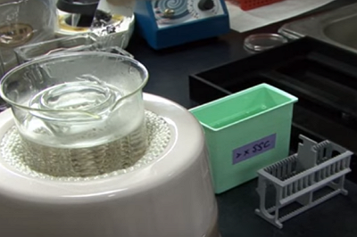
- 16.Remove rubber cement Slide into 2X SSC and remove cover glass2X SSC Room temp. 5 min2X SSC / 0.3% NP-40 73~75℃ 1-2min2X SSC Room temp. 1 min
- Counter stain
- 17.Apply 10μl DAPI Solution to target area*DAPI Paraffin embedded tissue 1500ng/ml
- 18.Put on cover glass Seal with manicure

- Examine


-
00:00
1.
Paraffin embedded tissue
-
00:03
1.1
Deparaffinized
-
00:23
1.2
Dehydrate
-
00:41
1.3
Air dry
-
00:52
1.4
Pre-treatment
-
01:24
1.5
Protease treatment
-
02:04
1.6
Dehydrate (Room temperature)
-
02:32
1.7
Air dry
-
02:34
2.
FISH protocol
-
02:36
2.1
Mark hybridizing area
-
02:42
2.2
Apply 10μl FISH probe for 22mm x 22mm area
-
02:54
2.3
Cover with cover glass and seal with rubber cement
-
03:27
2.4
Denature
-
03:36
3.
Incubation
-
03:55
4.
Remove rubber cement Slide into 2X SSC and remove cover glass
-
04:33
5.
Counter stain
-
04:35
5.1
Apply 10μl DAPI Solution to target area
-
04:43
5.2
Put on cover glass Seal with manicure
-
04:51
6.
Examine
- 位置
-
- 資料夾名稱
- Immunohistochemistry (免疫組織染色)
- 發表人
- 系統管理者
- 單位
- 賴亮全教授
- 建立
- 2018-12-03 11:24:29
- 最近修訂
- 2018-12-03 15:35:38
- 長度
- 05:01
- 引用
- 2
- 來源
- https://www.youtube.com/watch?v=dRmWZ3IUjFo